 Version
1.3
Version
1.3
 Package Name
com.imagery.imagerymedical
Package Name
com.imagery.imagerymedical
 Category
Sports and Health
Category
Sports and Health
 Size
453.8KB
Size
453.8KB

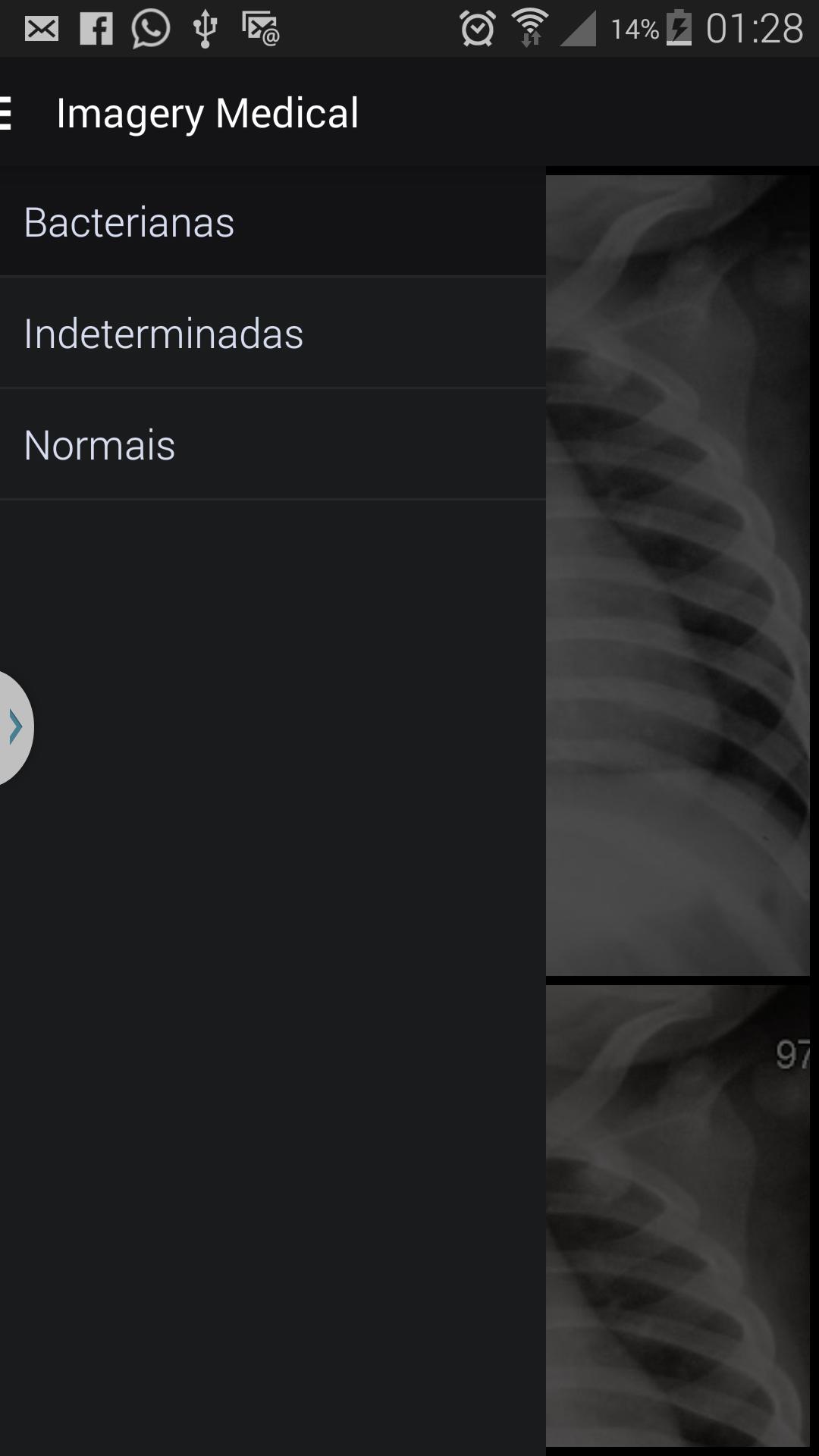
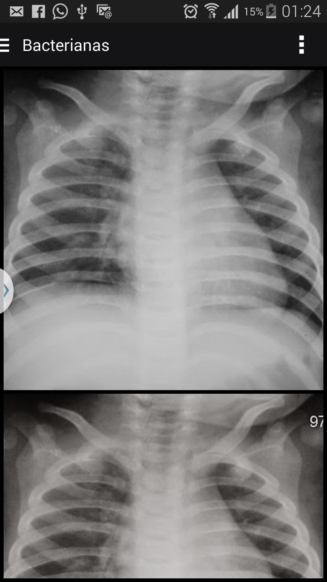
X-ray images to support the diagnosis of pneumonia in children
The relevance of pneumonia as a cause of child mortality in Brazil is duly proven in recent Ministry of Health documents. Pneumonia is the most important cause of childhood mortality, occupying the second cause of children's deaths in developing countries, including Brazil.
Chest X -ray is considered by the World Health Organization (WHO) as the best method currently available for the diagnosis of pneumonia in daily clinical practice. Studies on interobserving variation are common in all areas of medicine, however, predominate in image studies, given that in this area, the observer's performance represents the fragile part, contrasting to technological advancement, obtained in the last decade.
The interpretations that differ from a "consensus" by an experienced observer committee can be called "error." The variation between observers occurs when there is an error by an observer, but also includes cases where there is a difference of general opinion on what a correct interpretation represents. Errors and variations have been studied notably in the interpretation of radiographs.
For the determination of the validity of the results of any study, the accuracy of the diagnostic test should be as close as possible to reality, then the reference value, also called “standard
Gold ”Fundamental factor in the evaluation of diagnostic tests. In the case of radiographic diagnosis of the chest, especially in childhood, accuracy in image interpretation is subjectively evaluated through inter and intraobserving agreement, as there is rarely a pattern that can be used as a reference (standard) in the diagnosis of pneumonia.
Therefore, the current panorama of childhood pneumonia has the following characteristics:
(i) pneumonia are important cause of morbidity and mortality in childhood;
(ii) there is lack of diagnostic tests sensitive from the microbiological point of view;
(iii) highly effective childhood vaccines (HIB vaccines and antipneumococcal) are availability;
(IV) There is a lack of accurate diagnostic tests that are simple to execute and technological implementation, available in developing areas that may be subject to standardization to enable comparisons between studies. The advantages of using chest radiography in different
regions of the world in pneumonia surveillance studies justify their low cost; availability in most study sites; viability of comparison with other studies; Easy to digitize images, and storage; Possibility of standardization of readings and low iatrogenicity of the method.
Artificial Intelligence in Health The applications of expert systems using Artificial Intelligence in Medicine (AMI) are focused especially on
(i) alerts and reminders;
(ii) aid to the diagnosis;
(iii) therapeutic criticism;
(iv) information recovery agents; and
(v) recognition and interpretation of images. Among the current challenges for the implementation of AMI systems are the development of systems that accurately characterize aspects of medical practice.
In the last three decades computational vision (VC) comes as a new AI tool. VC based on processing images taking into account all the individual's individual knowledge and modeling this knowledge in the form of mathematical models and that aims to solve new problems based on previously solved problems. The important characteristic noted is the technological evolution that has as its product the standardization of the information coming from its application in the bulge of VC.


 Download APK(453.8KB)